University of Iowa researchers in engineering and medicine have won a five-year federal grant to significantly improve the resolution of brain scans, which could bolster the fight against diseases as people age.
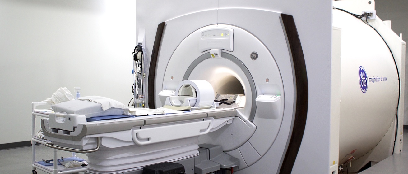
Sponsored by a National Institute on Aging grant, scholars believe they can generate ultra-high-resolution images of the brain through synergies in deep learning algorithms and novel sequences on a 7 Tesla (7T) Magnetic Resonance Imaging (MRI) scanner.
More accurate and detailed brain images would help physicians diagnose diseases earlier on when intervention could still make a difference, said Mathews Jacob, professor of electrical and computer engineering and an Iowa Technology Institute faculty affiliate who is principal investigator.
“The higher resolution enables us to detect and monitor changes early on so we can intervene, try new drugs, and more quickly monitor the impact of those drugs,” Jacob said.
High resolution brain scans would have a host of applications for people of all ages, he said.
The National Institute on Aging of the National Institutes of Health awarded the $3.75 million grant titled “Model Based Deep Learning Framework for Ultra-High-Resolution Multi-Contrast MRI” in December.
The grant incorporates the work of a multi-disciplinary team of researchers.
Model-based deep learning reconstruction algorithms developed in Jacob’s Computational Biomedical Imaging Group will work in combination with segmentation algorithms developed by co-investigator Xiaodong Wu, professor of electrical and computer engineering and an ITI faculty affiliate.
Co-investigators Merry Mani and Vincent Magnotta in the Department of Radiology will develop novel advanced multi-contrast sequences for use on the whole body 7T MRI scanner at the UI Magnetic Resonance Research Facility. The group closely collaborates with Iowa Institute of Neuroscience researchers Kumar Narayanan and Joel Geerling.
The goal of the research is to develop a 15‐minute 3D MRI scan that is not vulnerable to motion of the patient and through algorithms can accurately recover high resolution images of the entire brain. This process would solve a challenge with current MRI protocols in which patients struggle to remain still through lengthy scans leaving data susceptible to distortion.
Segmentation algorithms would allow physicians to inspect brain sub-structures, including hippocampal sublayers and cortical layers. The level of detail could enable screening of people at high risk for neurodegenerative disorders, determine early disease progression, and assess response to therapies.
This approach would also be considerably less expensive than positron emission tomography scans and does not involve radiation exposure.